"TISSUES" PART 2 (ANIMAL TISSUES)
TISSUES PART 2 (ANIMAL TISSUES)
Notes by:- Nagraj Sir
Animal Tissues are divided on the basis of function are four types:-
A.Epithelial Tissue
B. Muscular Tissue
C. Connective Tissue
D.Nervous Tissue
A.Epithelial Tissue:
the epithelial tissues are classified as follows:
- Squamous epithelium
- Cuboidal epithelium
- Columnar epithelium
- Glandular epithelium
- Ciliated epithelium
Differences between different types of epithelial tissues:
| S. No. | Characteristic property | Squamous epithelium | Cuboidal epithelium | Columnar or glandular epithelium | Ciliated epithelium |
| Shape of cells | It consists of thin, flat. Disc like polygonal or irregular-shaped cells with round and flat nucleus. | It consists of cube-like cells of almost equal height and width. | It consists of tall, cylindrical, pillar-like cells. Basal part of cell bears oval nucleus | It consists of tall cells with cytoplasmic hair like cilia at free ends. | |
| Appearance of cells | Adjacent cells fit together like tiles on a pavement or floor. | Cells appear square-like in vertical section but their free surface seems to be hexagonal. | The free end of the cells consists of finger-like projections called microvilli. | Cells may be cuboidal or columnar and are, therefore , also called ciliated cuboidal epithelium or ciliated columnar epithelium. | |
| Place of occurrence | Forms the lining of nose, pericardial cavity, blood vessels, lung alveoli etc. | Present in kidney tubules, salivary glands etc. | Present in the inner surface of stomach, intestine, gall bladder etc. | Present in the lining of trachea, fallopian tube, nasal passage etc. | |
| Functions |
|
|
|
|
B.Connective Tissue:
Types of connective tissue:
In animals, there are following five types of connective tissues:
- Areolar (loose) connective tissue
- Dense connective tissue
- Adipose connective tissue
- Skeletal tissue
- Fluid connective tissue
Aerolar (loose) connective tissue:
- It is a loose and cellular connective tissue. Its matrix consists of two kinds fibers: white collagen fibers and yellow elastic fibers.
- Aerolar connective tissue is found between the skin and muscles, around blood vessels and nerves and in the bone marrow.
- It fills the spaces between different tissues and organs, hence called packing tissue.
- It acts as supporting and packing tissue between organs lying in the body cavity.
- It helps in repair of tissues after an injury.

Dense connective tissue:
- It is a fibrous connective tissue. It is characterized by ordered and densely packed collection of fibers and cells.
- It is the chief 2 component of ligaments and tendons.
- 1.Ligaments: These are elastic structures made up of yellow elastic fibrous tissue s which connect bone to another. It has considerable strength. Ligaments contain very little matrix. Ligaments strengthen the joint and they permit normal movement but prevent over-flexing or over-extension. Sprain is caused by excessive pulling (stretching) of ligaments.
- 2.Tendons: Tendons are cord like, strong inelastic structures that join skeletal muscles to bones. They are composed of white collagen fibrous tissue.
- It has great strength but its flexibility is limited.
Adipose tissue:
- It consists of large number of oval and rounded adipose cells (adipocytes) filled with fat globules.
- The adipose tissue is abundant below the skin, between the internal organs (e.g., around the kidney) in yellow bone marrow.
- Adipose tissue acts as food reservoir by storing fat.
- It acts as an insulator and regulates body temperature.
Skeletal tissue:
- Skeletal connective tissue forms the endoskeleton of the body of vertebrates. It includes cartilage and bone.
S.No. Bone Cartilage 1. They are hard and flexible endoskeleton. They are soft and flexible endoskeleton. 2. Porous in nature. Non-porous in nature. 3. Blood vessels are present. Blood vessels are absent.
6. Bone cells are known as osteocytes. The cells in matrix are called chondriocytes.
8. Long bones contain bone marrow in hollow, narrow cavity. Bone marrow absent. Cartilage is always solid. 9. Bones are present in the whole body forming internal skeletal framework. Cartilage are present at the joints of bones, in external ear (pinna), nose tip, epiglottis, trachea etc. 10. Functions:
- It provides shape to the body.
- It provides skeletal support to body.
- It protects vital body organs such as brain, lungs etc.
Functions:
- Cartilage provides support and flexibility to the body parts.
- It smoothens body surfaces at joints.
- It is a loose and cellular connective tissue. Its matrix consists of two kinds fibers: white collagen fibers and yellow elastic fibers.
- Aerolar connective tissue is found between the skin and muscles, around blood vessels and nerves and in the bone marrow.
- It fills the spaces between different tissues and organs, hence called packing tissue.
- It acts as supporting and packing tissue between organs lying in the body cavity.
- It helps in repair of tissues after an injury.
Dense connective tissue:
- It is a fibrous connective tissue. It is characterized by ordered and densely packed collection of fibers and cells.
- It is the chief 2 component of ligaments and tendons.
- 1.Ligaments: These are elastic structures made up of yellow elastic fibrous tissue s which connect bone to another. It has considerable strength. Ligaments contain very little matrix. Ligaments strengthen the joint and they permit normal movement but prevent over-flexing or over-extension. Sprain is caused by excessive pulling (stretching) of ligaments.
- 2.Tendons: Tendons are cord like, strong inelastic structures that join skeletal muscles to bones. They are composed of white collagen fibrous tissue.
- It has great strength but its flexibility is limited.
Adipose tissue:
- It consists of large number of oval and rounded adipose cells (adipocytes) filled with fat globules.
- The adipose tissue is abundant below the skin, between the internal organs (e.g., around the kidney) in yellow bone marrow.
- Adipose tissue acts as food reservoir by storing fat.
- It acts as an insulator and regulates body temperature.
Skeletal tissue:
- Skeletal connective tissue forms the endoskeleton of the body of vertebrates. It includes cartilage and bone.
| S.No. | Bone | Cartilage |
| 1. | They are hard and flexible endoskeleton. | They are soft and flexible endoskeleton. |
| 2. | Porous in nature. | Non-porous in nature. |
| 3. | Blood vessels are present. | Blood vessels are absent. |
| 6. | Bone cells are known as osteocytes. | The cells in matrix are called chondriocytes. |
| 8. | Long bones contain bone marrow in hollow, narrow cavity. | Bone marrow absent. Cartilage is always solid. |
| 9. | Bones are present in the whole body forming internal skeletal framework. | Cartilage are present at the joints of bones, in external ear (pinna), nose tip, epiglottis, trachea etc. |
| 10. | Functions:
| Functions:
|
Fluid connective tissue: Fluid connective tissue links the different parts of the body and maintains continuity in the body. It includes blood and lymph.
Blood:
- Blood is fluid connective tissue. In this tissue cells move in a fluid or liquid matrix or medium called blood plasma.
- The blood plasma does not contain protein fibres but contain cells called blood corpuscles or blood cells. These blood corpuscles and cells are:
Red blood corpuscles (RBC) or erythrocytes
White blood corpuscles (WBC) or leucocytes
Platelets
RBCs and WBCs are living, while plasma and platelets are non-living.
C.Nervous tissue:
- A tissue which is specialized to transmit messages in our body is nervous tissue. Brain, spinal cord and nerves are all composed of nervous tissue.
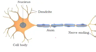
- Nervous tissue contains highly specialized unit cells called nerve cells or neurons.
- A neuron consists of a cell body (cyton or soma) with a nucleus and cytoplasm, from which long thin hair- like parts arise called dendrons.
- Dendrons further branched out to form dendrites. From the distal part of cyton arises a very long process called axon.
D. Muscular tissue:
- Striated muscles (stripped, skeletal or voluntary muscles)
- Smooth muscles (unstriated, visceral or involuntary muscles)
- Cardiac muscles
| S.No. | Unstriated muscles | Striated muscles | Cardiac muscles |
| 1. | Present in the wall of alimentary canal, blood vessels, respiractory tract, urinary bladder etc. | Present in limbs, tongue, body wall and pharynx. | They are present in the wall of heart and valve |
| 2. | Muscle fibres are spindle- shaped. | Muscle fibres are cylindrical. | Muscle fibres are cylindrical. |
| 3. | Fibres are unbranched. | Fibres are unbranched. | Fibres are branched. |
| Muscle cells are multinucleate. | Muscle cells are uninucleate. | Muscle cells are uninucleate or some time multinucleate | |
| Nerve supply from autonomous nervous system. | Nerve supply from central nervous system. | Nerve supply from both autonomous and central nervous system. | |
| Cross striations absent. | Dark and light bands (cross striations) present. | Cross striations and intercalated disc present. | |
| Involuntary. | Voluntary. | Involuntary. | |
| Do not get fatigued. | Get fatigued. | Do not get fatigued. | |
| Function: Cause contraction and mobility in visceral organs and involuntary muscles. | Function: Cause movement of limbs and locomotion. | Function: cause heartbeat. |
Comments
Post a Comment